Written by Dr. Roupen Odabashian MD, FRCPC
Oncologist | Founder, MeDucation AI | Updated October 2026
The verdict: disseminated intravascular coagulation (DIC) is not a single lab value, it is a clinical diagnosis built from two pieces: a predisposing condition known to trigger DIC, and a constellation of laboratory abnormalities that fits the picture[1]. The ISTH overt DIC score formalizes the second piece, a score of 5 or more is compatible with overt DIC, and the committee that built it was explicit that scoring should happen daily because the trend matters more than any single number[2]. Treatment has one cornerstone that outranks everything else: treat the underlying disorder, whether that is sepsis, an obstetric catastrophe, trauma, or acute promyelocytic leukemia (APL)[3]. Blood products are reserved for bleeding patients or those facing an invasive procedure, not for correcting numbers in isolation[3], and heparin has a narrow, specific role rather than a routine one[1][3].
Conflict of interest, up front: I am a practicing hematologist-oncologist and I founded MeDucation AI, which sells a physician-written heme/onc question bank and AI-powered study tools. Nothing below is written to sell you anything, this is a bedside and boards reference, and I link to the primary guidelines and scoring literature throughout.
Quick-reference summary
Question | Bottom line |
|---|---|
What defines DIC? | Intravascular activation of coagulation with loss of localization, causing microvascular injury and organ dysfunction[2] |
How is it diagnosed? | A predisposing condition plus a compatible lab pattern, formalized by the ISTH overt DIC score[2][4] |
What score confirms DIC? | 5 or more points is compatible with overt DIC, though the committee called this tentative pending further validation[2] |
Single most important treatment step? | Treat the underlying cause; everything else is supportive[3] |
When to transfuse platelets/plasma? | Generally reserved for active bleeding or a planned invasive procedure, not lab numbers alone[3] |
Is heparin routine? | No, reserved for clinical thrombosis, purpura fulminans, or another independent indication[1][3] |
Classic oncologic cause? | Acute promyelocytic leukemia, treated with early ATRA even before genetic confirmation[5][6] |
How do you calculate the ISTH DIC score?
The ISTH overt DIC score uses four lab values and only applies once you already suspect a DIC-associated condition[4]. The subcommittee was explicit that the algorithm requires "the presence of an underlying disorder known to be associated with DIC" as a precondition before scoring is even meaningful[2]. This matters on boards and at the bedside: you do not apply the score to an isolated thrombocytopenic patient with no plausible trigger.
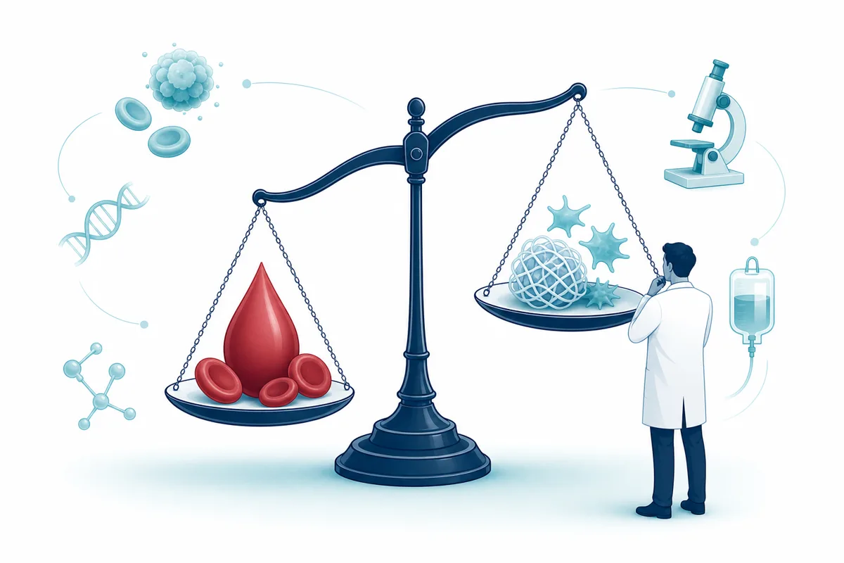
Component | Result | Points |
|---|---|---|
Platelet count | Values are weighted by severity of thrombocytopenia (lower counts score higher); the JSTH criteria score ≤50 ×10³/µL as 3 points, 51 to 80 as 2, and 80 to 120 as 1[7] | 0 to 3 |
Fibrin-related marker (D-dimer or FDP) | D-dimer is "invariably elevated in DIC, often dramatically (e.g., >4,000 ng/mL)"; a normal D-dimer essentially excludes DIC[1] | 0 to 3 |
Prolonged prothrombin time | PT elevation supports DIC but can be normal in over half of patients[1] | 0 to 2 |
Fibrinogen level | Fibrinogen is "the least sensitive marker of DIC, with a sensitivity of only 25%"; sepsis can elevate it independently, so a normal level does not rule DIC out[1] | 0 to 1 |
A total score of 5 or more is "compatible with DIC," while a lower score "may be indicative (but is not affirmative) for non-overt DIC"[2]. The ISTH subcommittee recommended daily rescoring to track severity and course[2], and emergency medicine literature echoes this: a fibrinogen "actively falling from 300 mg/dL to 100 mg/dL is more worrisome than a fibrinogen that is stably low at 100 mg/dL"[1]. The score has been prospectively validated in critically ill patients[7], and the British Society for Haematology (BSH) guideline states plainly that the ISTH score "provides objective measurement of DIC" and correlates with clinical outcomes when DIC is present[3].
How do you separate DIC from TTP, HIT and liver failure?
DIC is a clinical diagnosis made by combining a trigger with a lab pattern and excluding mimics[1]; the mimics are where fellows get tripped up on rounds and on boards. Each of the major look-alikes has a clean distinguishing feature once you know where to look.
Feature | DIC | TTP/HUS | HIT (alone) | Cirrhosis/liver failure |
|---|---|---|---|---|
Coagulation factors (PT, PTT, fibrinogen) | Often abnormal; PT/PTT can be normal in many cases of DIC[1] | Normal, "clots are composed of platelets and don't involve coagulation system activation"[1] | Normal INR and fibrinogen[1] | Chronically elevated INR, often stable at baseline[1] |
Platelet count | Moderate thrombocytopenia, platelets below 30,000/µL are uncommon[1] | Often profound, with exuberant schistocytosis[1] | Not usually profoundly reduced (platelets often >20,000/µL) unless DIC is superimposed[1] | Can be low but typically stable over time[1] |
Factor VIII | Low (consumed)[1] | Not a distinguishing feature | Not a distinguishing feature | Normal or elevated, since it is made by endothelial cells, not hepatocytes[1] |
Key clinical clue | Occurs gradually around the time of the precipitating illness[1] | Fever with thrombocytopenia and MAHA, distinct from a trauma/sepsis trigger[7] | Abrupt onset, beginning after heparin exposure[1] | Stable, chronic pattern that tracks with known liver disease, not an acute trigger[1] |
Two further points are worth holding onto. First, HIT and DIC are not mutually exclusive, severe HIT can trigger DIC in roughly 10% of cases, and when it does, platelets fall more profoundly and INR/fibrinogen abnormalities appear[1]. Second, cirrhosis is itself a risk factor for DIC because baseline depletion of procoagulant and anticoagulant proteins makes the system easier to tip over, and advanced cirrhosis can also cause a primary hyperfibrinolytic state called accelerated intravascular coagulation and fibrinolysis (AICF) that looks similar[1]. Thrombotic microangiopathies (TTP, HUS) are distinguished by a fundamentally different mechanism, thrombosis there is driven by platelet activation via ADAMTS13 deficiency and ultralarge von Willebrand factor rather than tissue factor-driven coagulation activation, so coagulation assays stay normal[7].
What is a good mnemonic for the causes of DIC?
There is no single verified mnemonic in the sources reviewed here, but the causes do sort cleanly into categories worth memorizing as a checklist rather than forcing an acronym. Infection (particularly gram-negative sepsis and toxigenic organisms), malignancy (acute promyelocytic leukemia and monocytic leukemia causing bleeding-predominant DIC, adenocarcinomas of the pancreas, prostate, lung, gastric and ovarian origin causing a chronic, thrombosis-predominant form), obstetric catastrophe (abruption, amniotic fluid embolism, HELLP, retained dead fetus, septic abortion), trauma and direct tissue damage (burns, traumatic brain injury, fat embolism), and a miscellaneous bucket covering severe hepatitis, vascular malformations, catastrophic antiphospholipid syndrome, and HLH[1][7]. Acute DIC causes abrupt, exuberant factor depletion (think sepsis), while chronic DIC, typically from disseminated adenocarcinoma, causes gradual consumption that the body can partly compensate for, producing a milder, "chronic compensated" picture[1].
When should you give platelets, cryoprecipitate or plasma?
Transfuse for bleeding or an impending procedure, not to chase a number. The BSH guideline is explicit: "Transfusion of platelets or plasma (components) in patients with DIC should not primarily be based on laboratory results and should in general be reserved for patients who present with bleeding"[3]. For non-bleeding patients, prophylactic platelet transfusion is not given unless there is a perceived high bleeding risk[3].
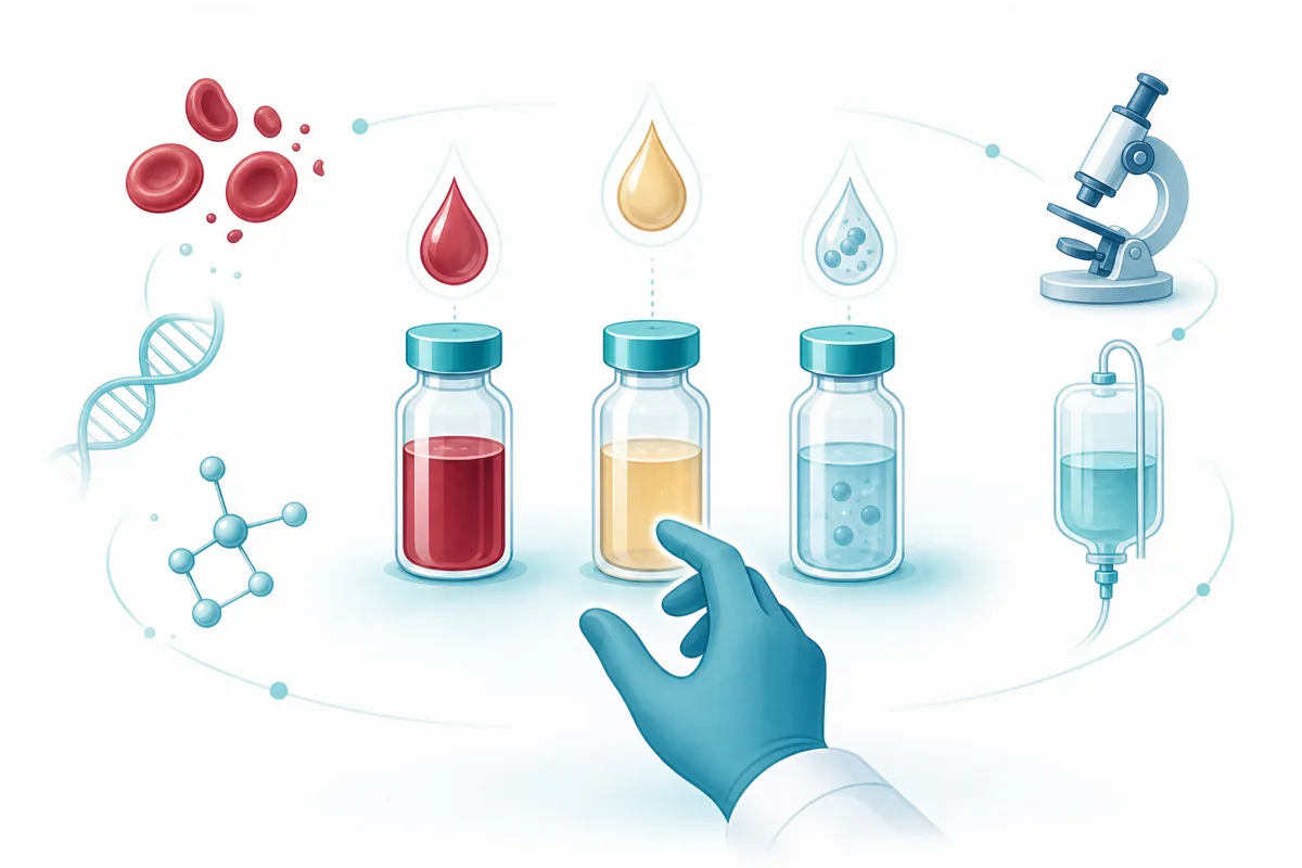
Product | Threshold / target | Source |
|---|---|---|
Fresh frozen plasma (FFP) | Consider in bleeding patients with prolonged PT/aPTT, or before an invasive procedure; recommended when fibrinogen is below 100 mg/dL in actively bleeding patients; not based on labs alone | [3][8] |
Fibrinogen (cryoprecipitate or concentrate) | Reasonable to keep fibrinogen above roughly 50-100 mg/dL generally, and above roughly 150 mg/dL for actively bleeding patients or those facing a procedure; severe hypofibrinogenemia (<1 g/L) persisting despite FFP may need cryoprecipitate or fibrinogen concentrate | [1][3] |
Because ongoing consumption continues to eat platelets, results from transfusion can be "disappointing," which is a useful phrase to remember for why repeat transfusion sometimes feels futile without treating the underlying trigger[1]. Washed packed red cells and platelet concentrates are generally considered safe in uncontrolled DIC, while specialized products like cryoprecipitate and FFP can meaningfully alleviate the coagulopathy when targeted correctly[8].
Is heparin ever used in DIC?
Yes, but narrowly, not as a routine therapy. "Unfortunately, research into therapeutic heparin anticoagulation has generally been unimpressive. Thus, heparin anticoagulation isn't indicated in most cases of DIC"[1]. The specific situations where it is reasonable include established clinical thrombosis such as DVT or pulmonary embolism, purpura fulminans (which requires its own distinct management approach), severe digital ischemia, and cases where another independent indication for anticoagulation exists, such as atrial fibrillation[1]. The BSH guideline agrees: therapeutic-dose heparin should be considered specifically "where thrombosis predominates, such as arterial or venous thromboembolism, severe purpura fulminans associated with acral ischemia or vascular skin infarction"[3].
Dosing is complicated by heparin resistance in DIC, because low antithrombin-III levels blunt heparin's effect, meaning higher doses than usual may be required, and weight-based low molecular weight heparin without anti-Xa monitoring risks underdosing[1]. Continuous infusion unfractionated heparin may be preferred in patients with a coexisting high bleeding risk because of its short half-life and reversibility[3]. Separately, for critically ill, non-bleeding DIC patients, prophylactic-dose heparin or LMWH for VTE prevention is recommended, which is a different question from therapeutic anticoagulation[3].
Why are antifibrinolytics usually avoided in DIC?
Because DIC can swing toward thrombosis as easily as it swings toward bleeding, and blocking fibrinolysis in a thrombosing patient is dangerous. "In general, antifibrinolytic agents should be avoided in DIC because they are known to produce thrombotic complications, such as myocardial infarction and renal artery thrombosis when there is systemic clotting"[8]. The BSH guideline reinforces this: "In general, patients with DIC should not be treated with antifibrinolytic agents"[3].
There is a narrow exception. Patients whose DIC is characterized by a primary hyperfibrinolytic state with severe bleeding could be treated with lysine analogues such as tranexamic acid[3], and antifibrinolytics may have a specific role in DIC driven by hyperfibrinolysis in acute promyelocytic leukemia and other cancers where alpha-2-antiplasmin is uniquely depleted[8]. Even in APL specifically, available trial evidence does not support routine tranexamic acid use, reserving it for life-threatening hemorrhage not responding to conventional therapy[5].
Why is acute promyelocytic leukemia the classic oncologic cause of DIC?
Because APL causes DIC through a uniquely aggressive, two-pronged mechanism, and because the definitive treatment of the coagulopathy is the leukemia-directed therapy itself, not blood products alone. Tissue factor on the surface of leukemic promyelocytes directly activates the coagulation cascade, while annexin II on the same cells activates tPA and uPA, driving hyperfibrinolysis that is "probably the primary driver of bleeding"[5].
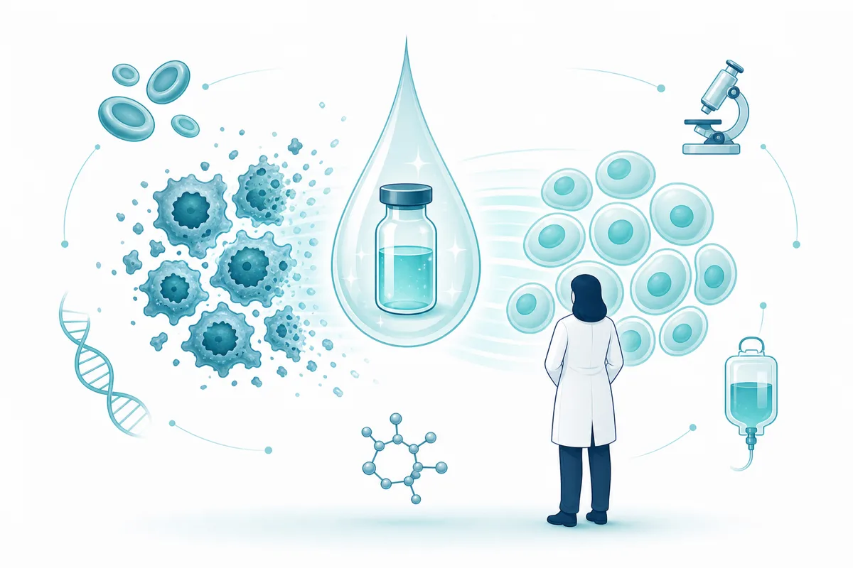
Nearly 90% of APL patients have some clinical bleeding at presentation, and intracranial and pulmonary hemorrhage account for most hemorrhagic deaths[5]. DIC, differentiation syndrome, and sepsis together drive the "early death" phenomenon, with around 20% of patients dying soon after presentation in some series, even though APL is highly curable, around 90%, once a patient survives the early period[5]. This is why all-trans retinoic acid (ATRA) should be started as soon as APL is clinically suspected, even before cytogenetic confirmation, ideally from the emergency department[5][6]. ATRA differentiates the malignant promyelocytes into neutrophils, which directly eliminates the DIC rather than just treating its downstream effects[5]. Supportive transfusion targets in APL are more aggressive than in other causes of DIC: platelet transfusions to maintain the platelet count at about 20,000/µL and cryoprecipitate to maintain a fibrinogen level at least above 100-150 mg/dL[6]. Heparin should generally be avoided in APL-associated DIC unless there is a severe thrombotic event, and its use has been associated with increased hemorrhagic events in this specific population[5].
For fellows building a broader study plan around high-yield hematologic emergencies like this one, our guide on building a 6-month study plan for the ABIM hematology and oncology boards walks through how to sequence topics like DIC and APL against the rest of the blueprint, and our piece on comparing heme/onc question banks can help you decide where to get volume once the concepts are solid.
FAQ
Is DIC diagnosed by a single lab test?
No. DIC rests on three components together: a compatible constellation of lab abnormalities, a known underlying disorder, and exclusion of an alternative explanation[1]. No single test, including D-dimer, is diagnostic on its own[1].
What ISTH score is needed to diagnose overt DIC?
A score of 5 or more is compatible with overt DIC, while a lower score may suggest non-overt DIC without confirming it, and the original committee described this cutoff as tentative, pending further prospective validation[2].
Does a normal PT or PTT rule out DIC?
No. INR and PTT abnormalities support the diagnosis but "can be normal in over half of patients with DIC," because these tests measure clotting factors only, not deficiencies in anticoagulant proteins like protein C and S[1].
Should DIC labs be checked once or serially?
Serially. The ISTH subcommittee recommends scoring daily to characterize severity and course[2], and the trend, a falling fibrinogen or a rapidly rising D-dimer, is often more informative than a single snapshot[1].
Is COVID-19 coagulopathy the same as DIC?
Not typically. COVID-19 coagulopathy is more prothrombotic and localized, most patients do not meet ISTH criteria for overt DIC, and the pattern, including only mild thrombocytopenia and sometimes a shortened rather than prolonged PT, differs from classical septic DIC[7].